Glycosaminoglycans


What gives the tensile strength and the resistance for compressional forces observed in Cartilages, Tendons and Ligaments? What maintains the shape of the Eye? From constituting heart valves to maintaining the flexibility of skin …. what is involved?  LETS FIND OUT.

There are  4 basic types of tissues, 

  1. Epithelial Tissue - are tissues that line surfaces found within animals
  2. Muscle Tissue - are groups of cells that construct the muscles of animals
  3. Nerve Tissue - are responsible for building up the nervous system
  4. Connective Tissue - is responsible in maintaining and supporting all other tissues

The spotlight of this article  goes to Connective Tissue. As this is a type of tissue that has the ability to connect other tissues and maintain their structure and function and even provide them with the nutrition needed. 

Connective tissue is composed of two components, Cells and the Extra-Cellular Matrix (ECM) which is secreated by the cells. The ECM is composed of 4 components they are,

  1. Fibres
  2. Multi-adhesive proteins
  3. Proteoglycans
  4. Glycosaminoglycans

The last 3 together is referred to as the Ground substance. This is where you find Glycosaminoglycans.

So, what is so special about these Glycosaminoglycans?

Glycosaminoglycans are heteropolysaccharides which are linear and unbranched. They are basically made of 2 sugar residues, 

  1. Amino sugars
  2. Uronic acid sugars

Two types of amino sugars can be found, Glucosamine and Galactosamine. Two types of Uronic acids can be found, Glucuronic acid and Iduronic acid. One from each bond together through glycosidic bonds to form the repeating disaccharide unit of the Glycosaminoglycan chain.

The other property of Glycosaminoglycans is its high negative density due to the presence of negative charges bonded to either of the sugar residues forming the disaccharide unit. This is responsible in attracting water molecules as a result glycosaminoglycans are found to be highly hydrated. This what gives the ground substance its gel like property.

Glycosaminoglycans (GAGs) are found in mainly in animals. There are mainly 6 GAGs that involve in making the Ground substance. Except one all the other 5 GAGs undergo Post-translational modifications where it bonds with a core protein forming Proteoglycans.

Hyaluronic acid is a GAG and deserves its own article but basically its the largest GAG and the one which doesn't bond with a core protein to form a proteoglycan. It involves in forming Proteoglycan aggregates.

The repeating disaccharide unit is formed of a Glucuronic acid and a N-acetylglucosamine compound bonded together by a 𝛃(1-3) glycosidic bond and each of the units are bonded by a 𝛃(1-4) glycosidic bond. 

It is found in the Synovial fluid of joints, cartilages, tendons, and vitreous humour of eye.

Chondroitin Sulfate is the most abundant GAG and it is mainly found in the Cartilages, Tendons, Ligaments and Aorta as well. Chondroitin sulfate is of 2 types,

  1. Chondroitin-4-Sulfate - it is composed of a Glucuronic acid and a N-acetylgalactosamine bonded together by a π›ƒ(1-3) glycosidic bond to form the repeating disaccharide units which are bonded together by π›ƒ(1-4) glycosidic bond. The 2 functional groups that renders this molecule negative are the Carboxylic acid group bonded to the 5th C of the Glucuronic acid and Sulfate group bonded to the 4th C of the N-acetylgalactosamine compound.
  2. Chondroitin-6-Sulfate - it has a similar structure to Chondroitin-4-Sulfate with the only difference being that the sulfate group is bonded to the 6th C of the N-acetylgalactosamine sugar residue.
Chondroitin-4-Sulfate is found primarily in growing cartilages and equal amounts of both Chondroitin-6-Sulfate and Chondroitin-4-Sulfate can be found in adult cartilages.

Chondroitin Sulfate also can be found found in the Central Nervous System where it contributes to the structural function in addition it involves with the signaling molecules which prevent the repair of damaged nerve endings.


Dermatan Sulfate is found in skin and involves in the pliability of the skin. In addition it is found in the blood vessels and heart valves. It has the same structure as Chondroitin-4-Sulfate except that the Uronic acid is a Iduronic acid which is a C5 epimer of the Glucuronic acid. This conversion is catalyzed by an epimerase enzyme.

Therefore the Glycosidic bond involved in forming the repeating Disaccharide unit is a 𝛂(1-3) bond. ( Curious as to why this is so? Check my article on Monosaccharides)


Keratan Sulfate is a GAG which was 1st extracted from the Cornea ( this is called Keratan Sulfate  or KS Ⅰ ). This is responsible for the transparency of the cornea. In the absence of KS Ⅰ opacity of cornea is observed. KS Ⅰ was found in the Cartilage of animals in addition it is also found in horny parts such as hoofs, hair, nails and horns.

KS is special in that it lacks a Uronic acid like other GAGs. Instead it has a Galactose compund bonded to a N-acetylglucosamine compound. But then where does it get its negative charge density? the sulfate group bonded to the 6th C renders it negative.

A π›ƒ(1-4) glycosidic bond  is involved in forming the repeating disacchride unit. Each unit is then bonded together by a 𝛃(1-3) glycosidic bond.


Heparin is not found as an Extra-cellular component but rather as an Intra-cellular component which is synthesized and secreated by the Mast cells found in the Extra-cellular matrix of Connective tissue. It is acts as an anti-coagulant by binding with anti thrombin to inhibit the formation of thrombin, factor XⅠ and factor ⅠX.

It has the highest negative charge density that is each disaccharide unit has 2.5 sulfate groups each. The other structural difference that we observe compared to other GAGs is that the glucosamine sugar lacks a acetyl group. Instead it has a N-sulfate group.

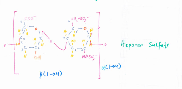
The majority of the uronic acids during the beginning of the synthesis of the heparin chain is Glucuronic acid but by the completion 90% of them are converted into Iduronic acids which is catalyzed by an epimerase enzyme. The glycosidic bond involved in forming the disaccharide unit and bonding each unit together is a π›‚(1-4) bond.


Heparan Sulfate is similar to heparin but it has a lower negative charge density. It is composed of lesser sulfate groups and instead it has carboxylic groups in addition the amino sugar is Non-acetylated. The majority of the uronic acids involved are Glucuronic acids. Therefore compared to heparin a 𝛃( 1-4 ) glycosidic  bond is involved in forming the disaccharide unit and each unit bonded together by π›‚(1-4) glycosidic bonds.

A prominent function of HS is wound repair. In addition it is a membrane component where it is found attached to protein complexes forming proteoglcans and they involve in maintaining cell growth, cell proliferation and cell to cell communication. It is also found in the basement membrane of the kidney and involves in selective filtration of metabolites during Glomerular filtration.


Comments

Popular posts from this blog

Carbohydrates 1 - Monosaccharides

What is Sex Linkage?

4 Major Biological Molecules - Carbohydrates

Essential Elements

Carbohydrates 2 - Disaccharides Ⅰ

Carbohydrates 2 - Disaccharides Ⅱ

Carbohydrates 3 - Polysaccharides

Hyaluronic acid